

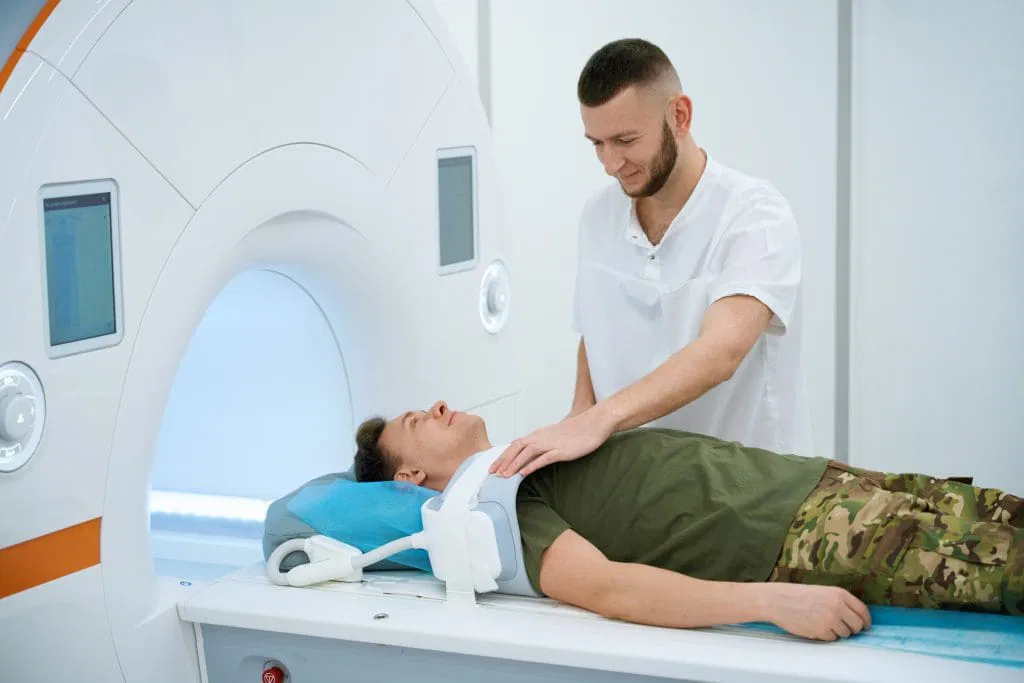
Nuclear medicine is a highly specialized branch of radiology that uses small doses of radioactive materials in the body to help to diagnose internal conditions. Doctors use radioactive tracers to observe internal processes, and they track the distribution of the material over a testing period. Because these tracers emit measurable radiation from within the patient, specialized scanners can detect the signals to generate highly detailed images of internal structures. These procedures are safe and monitored by trained doctors and staff.
How It Works
For nuclear medicine testing, healthcare providers may inject a specific radiotracer into a patient, and the substance travels through the circulatory system to a specific organ. As the specialized tracer circulates, the radioactive compounds naturally accumulate in certain structures, which can include the heart. A specialized camera captures the resulting radiation rays from multiple angles around the patient.
When the scanning equipment gathers the radiation data, a computer processes the data into a visual representation of the heart. The diagnostic images display patterns of absorption within heart tissue and arteries. Certain levels of radiotracer absorption indicate healthy blood flow through the heart and major blood vessels, so deviations from these standards can suggest improper levels of circulation.
What It Shows
Advanced nuclear imaging techniques measure the amount of blood reaching heart tissue during exercise and during rest. If a patient undergoes a stress test, the physician can compare the scan results against the established baseline images. A difference between these two states highlights potential blockages, and the medical team uses this data to quantify cardiovascular risk.
This procedure can identify scar tissue from prior cardiac events, and it maps the location of any damaged heart muscle. The overall status is evaluated by a cardiologist to determine the extent of potential disease. Although some areas might display reduced blood supply, other segments of the heart muscle might function normally during the examination.
Because these diagnostic tests assess cellular activity, they can reveal early physiological signs of heart disease before symptoms fully manifest. Medical professionals measure how much blood pumps through the heart, and this calculation shows the overall efficiency of the heart. This quantitative metric guides future treatment plans for caring for the patient.
How It’s Different
Traditional imaging methods like echocardiograms primarily display the basic structure of the heart valves and the muscular walls. While a standard angiogram outlines the interior shape of the arteries, a nuclear medicine scan measures the actual function of the cardiac tissues. Conventional electrocardiograms only record superficial electrical signals, but advanced nuclear cardiology tracks the deeper cellular function and blood flow. These images are helpful for finding smaller issues that traditional testing may not be able to catch.
Schedule a Nuclear Medicine Appointment
Patients who have found that other testing methods for heart health are not aiding in the diagnostic process, nuclear medicine diagnostic testing may be helpful in creating a treatment plan. Physicians who use this method can catch heart problems early, and patients can receive earlier treatment. Schedule an appointment with a radiology specialist to begin nuclear medicine testing.
